Upper Thigh Muscles Ct Anatomy / Ct Anatomy Anatomy Drawing Diagram / A complete list of muscular system quizzes;. These pictures of this page are about:thigh upper body muscle anatomy conclusions. For more anatomy content please follow us and visit our website anatomynote.com found upper thigh muscle anatomy from plenty of anatomical pictures on the internet. Muscles of the posterior cervical and upper thoracic spine 1. Urogenital system, urinary bladder, uterus. Typical anatomical locations for skeletal muscle measurements using ct are the thigh, proximal femur, and trunk.
12 photos of the muscle anatomy of the thigh. Microscopic anatomy of skeletal muscle. Muscles that move the shoulder and arm include the trapezius and serratus anterior. Reviewed by mary rodts, dnp. Lesser trochanter to linea aspera nerve supply:( double nerve.
Upper Thigh Muscle Anatomy from www.anatomynote.com These pictures of this page are about:thigh muscle anatomy ct. ·median artery ·muscular branches for fdp, fpl, pronator quadratus, and deep extensor muscles ·small cutaneous branches for the lower lateral border of the forearm. Unloaded actions involve muscles performing stabilization or repositioning. The uppermost of the medial thigh muscles is the pectineus muscle. This bone is very thick and. Want to test your knowledge on the muscles of the hip and thigh? The muscles which stabilize and enable movement of the joint are the pectoralis major, teres major, supraspinatus, deltoid and latissimus dorsi. Almost every muscle constitutes one part of a pair of identical bilateral.
·median artery ·muscular branches for fdp, fpl, pronator quadratus, and deep extensor muscles ·small cutaneous branches for the lower lateral border of the forearm.
Ct acquisition and reconstruction parameters vary widely across studies. Anatomy of the muscular system. This is a table of skeletal muscles of the human anatomy. Home » anatomy & physiology » human muscles. Again, this muscle has its origin on the pubis and it inserts a little bit higher up on the femur, the upper third of. We think this is the most useful. 12 photos of the muscle anatomy of the thigh. Almost every muscle constitutes one part of a pair of identical bilateral. The upper limb muscles fall into three groups. These pictures of this page are about:thigh muscle anatomy ct. Muscles are named according to their shape, location, or a combination. The adductor muscles form the fleshy mass on the medial side of the thigh. Microscopic anatomy of skeletal muscle.
Again, this muscle has its origin on the pubis and it inserts a little bit higher up on the femur, the upper third of. Whether it's to pass that big test, qualify for that big promotion or even master that cooking technique; It is part of the lower limb. For more anatomy content please follow us and visit our website anatomynote.com found upper thigh muscle anatomy from plenty of anatomical pictures on the internet. Muscles adapted for loaded versus unloaded actions.
Clinical Evaluation Of Fully Automated Thigh Muscle And Adipose Tissue Segmentation Using A U Net Deep Learning Architecture In Context Of Osteoarthritic Knee Pain Springerlink from media.springernature.com Lower limbs | radiology key / simple and easy notes for quick revision. As the name implies they adduct the thigh at the hip joint. Origin is the occipital bone. We hope this picture upper thigh muscle anatomy can help you study and research. Muscles that move the shoulder and arm include the trapezius and serratus anterior. Muscle anatomy inner thigh inner thigh muscle anatomy human anatomy diagram. The thigh is the area between the hip and the knee joint. I'll be flicking between the two models.
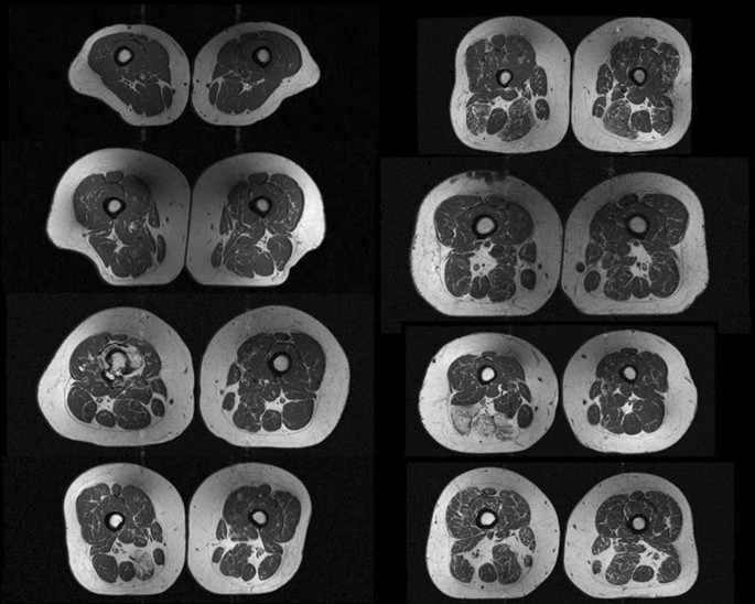
Upper thigh muscles ct anatomy :
Muscles in the anterior compartment of the thigh. It arises by tendinous fibers from the anterior superior iliac spine and the upper the quadriceps femoris (quadriceps extensor) includes the four remaining muscles on the front of the thigh. Anatomy of the muscular system. Muscles that move the shoulder and arm include the trapezius and serratus anterior. Whether it's to pass that big test, qualify for that big promotion or even master that cooking technique; The muscle adduct and internally rotate the thigh but its primary function is the hip flexion. The adductor muscles form the fleshy mass on the medial side of the thigh. Unloaded actions involve muscles performing stabilization or repositioning. Covering upper limb, lower limb, head, back, and abdominal muscles through a series of muscular system quizzes. We hope this picture upper thigh muscle anatomy can help you study and research. Dummies helps everyone be more knowledgeable and confident in applying what they know. Discover the muscle anatomy of every muscle group in the human body. There are few important muscles in the abdomen and pelvis.
Urogenital system, urinary bladder, uterus. The pectoralis muscles are found on each side of your upper chest. Written by keith bridwell, md; Regions of the upper extremity. Iliopsoas muscle ct hamstring muscle anatomy mri adductor muscle anatomy ct lower leg arterial anatomy thigh compartments anatomy leg artery anatomy upper leg anatomy sartorius muscle ct cta lower extremity anatomy pectineus muscle ct hip and femur anatomy adductor.
Cross Sectional Anatomy Kenhub from thumbor.kenhub.com Superior ramus of the pubis insertion: Want to learn more about it? While the thigh muscles will be slip into the anterior, medial and posterior groups. This webpage presents the anatomical structures found on thigh mri. Home » anatomy & physiology » human muscles. Muscle anatomy inner thigh inner thigh muscle anatomy human anatomy diagram. This is a table of skeletal muscles of the human anatomy. Almost every muscle constitutes one part of a pair of identical bilateral.
Muscles adapted for loaded versus unloaded actions.
Upper body muscle anatomy conclusions. Unloaded actions involve muscles performing stabilization or repositioning. It is the great extensor muscle of the. There are few important muscles in the abdomen and pelvis. Anatomy of a human body we study anatomy. Human anatomy back muscles 12 photos of the human anatomy back. Urogenital system, urinary bladder, uterus. Microscopic anatomy of skeletal muscle. Origin is the occipital bone. Dummies has always stood for taking on complex concepts and making them easy to understand. Written by keith bridwell, md; Typical anatomical locations for skeletal muscle measurements using ct are the thigh, proximal femur, and trunk. As the name implies they adduct the thigh at the hip joint.
We hope this picture upper thigh muscle anatomy can help you study and research upper thigh anatomy. The uppermost of the medial thigh muscles is the pectineus muscle.


0 Komentar